Human Bladder Cancer Antigen (UBC) Enzyme Linked Assay ELISA Kit Instructions for Use This reagent is for research use only. Specimen: serum or plasma Shanghai Qiao Yu Biological Professional Supply: Purpose of use: This kit is used to determine the content of bladder cancer antigen (UBC) in human serum, plasma and related liquid samples. Test principle: The UBC kit is a solid phase sandwich enzyme-linked immunosorbent assay (ELISA). Standards of UBC concentration and samples of unknown concentration are added to the microplates for detection. UBC and biotinylated antibodies are first incubated simultaneously. After washing, avidin-labeled HRP was added. After incubation and washing, the unbound enzyme conjugate is removed, then the substrates A, B, and the enzyme conjugate are added simultaneously. Produce color. The depth of the color is proportional to the concentration of UBC in the sample. Kit content and its preparation Kit components (2-8 ° C) 96-well configuration 48 hole configuration Formulation 96/48 ELISA plate 1 board (96T) Half board (48T) Ready-to-use Plastic diaphragm cover 1 block Half block Ready-to-use Standard: 80ng/ml 1 bottle (0.6ml) 1 bottle (0.3ml) According to the instructions for thinning Blank control 1 bottle (1.0ml) 1 bottle (0.5ml) Ready-to-use Standard dilution buffer 1 bottle (5ml) 1 bottle (2.5ml) Ready-to-use Biotinylated anti-UBC antibody 1 bottle (6ml) 1 bottle (3.0ml) Ready-to-use Affinity chain enzyme-HRP 1 bottle (10ml) 1 bottle (5.0ml) Ready-to-use Washing buffer 1 bottle (20ml) 1 bottle (10ml) Dilute according to the instructions Substrate A 1 bottle (6.0ml) 1 bottle (3.0ml) Ready-to-use Substrate B 1 bottle (6.0ml) 1 bottle (3.0ml) Ready-to-use Stop solution 1 bottle (6.0ml) 1 bottle (3.0ml) Ready-to-use Specimen dilution 1 bottle (12ml) 1 bottle (6.0ml) Ready-to-use Self-contained materials 1. Distilled water. 2. Sampler: 5ul, 10ul, 50ul, 100ul, 200ul, 500ul, 1000ul. 3. Oscillators and magnetic stirrers, etc. safety 1. Avoid direct contact with the stop solution and substrates A, B. Once exposed to these liquids, rinse with water as soon as possible. 2. Do not eat, drink, smoke or use cosmetics during the experiment. 3. Do not use your mouth to take any ingredients from the kit. Operational precautions 1. Reagents should be stored according to the label instructions and returned to room temperature before use. Standards after dilution should be discarded and cannot be stored. 2. The slats not used in the experiment should be immediately put back into the bag and sealed to prevent deterioration. 3. Other reagents not used should be packaged or covered. Do not mix reagents of different batches. Use before warranty. 4. Use a disposable tip to avoid cross-contamination. Avoid pipettes with metal parts when drawing stop solution and substrate A and B. 5. Use a clean plastic container to configure the wash solution. Mix all the ingredients and samples in the kit thoroughly before use. 6. Wash the enzyme plate should be fully patted dry, do not put the absorbent paper directly into the enzyme standard reaction well to absorb water. 7. Substrate A should be volatilized to avoid opening the lid for extended periods of time. Substrate B is sensitive to light and avoids prolonged exposure to light. Avoid contact with hands and be toxic. The OD value should be read immediately after the experiment is completed. 8. The order of addition of reagents should be consistent to ensure that all wells are incubated for the same time. 9. The incubation was carried out according to the time indicated in the instructions, the amount of addition and the order. Sample collection, processing and storage methods 1. Serum ---- Avoid any cell irritation during the procedure. Use tubes without pyrogens and endotoxins. After collecting the blood, the serum and red blood cells were quickly and carefully separated by centrifugation at 1000 x g for 10 minutes. 2, plasma ----- EDTA, citrate, heparin plasma can be used for testing. The pellet was removed by centrifugation at 1000 x g for 30 minutes. 3. Cell supernatant - 1000 x g was centrifuged for 10 minutes to remove particles and polymer. 4, tissue homogenate ----- the tissue is added to the appropriate amount of physiological saline mash. Centrifuge at 1000 × g for 10 minutes, take the supernatant 5. Storage ------If the sample is not used immediately, it should be divided into small parts - 70 °C to avoid repeated freezing. Do not use hemolysis or hyperlipemia as much as possible. If there are large amounts of particles in the serum, centrifuge or filter before testing. Do not heat thaw at 37 ° C or higher. It should be thawed at room temperature and ensure that the sample is fully thawed evenly. Reagent preparation 1. Standard: The serial dilution of the standard should be prepared during the experiment and cannot be stored. Mix the standard shakes before dilution. The dilution ratio is as follows: 80 ng/ml (No. 6 standard) The original concentration was directly added to 50 ul without dilution. 40 ng/ml (No. 5 standard) Add 100 ul of standard dilution to 100 ul of the original standard 20 ng/ml (No. 4 standard) 100ul of standard 5 is added to 100ul of standard dilution 10 ng/ml (No. 3 standard) 100ul of standard 4 is added to 100ul of standard dilution 5.0 ng/ml (No. 2 standard) 100ul of standard 3 is added to 100ul of standard dilution 2.5 ng/ml (No. 1 standard) 100ul of standard 2 is added to 100ul of standard dilution 0 ng/ml (blank control) The original concentration was directly added to 50 ul without dilution. 2. Dilution of wash buffer (50 x): 50-fold dilution of distilled water. Steps 1. Mix all reagents thoroughly before use. Do not allow the liquid to generate a large amount of foam, so as to avoid adding a large amount of air bubbles during the loading, resulting in errors in the loading. 2. The number of slats required is determined by the number of samples to be tested plus the number of standards. Multiple holes are recommended for each standard and blank hole. Each sample is determined according to its own quantity, and it is possible to use the double hole as much as possible to make a double hole. The specimen was diluted 1:1 with the specimen dilution and 50 ul was added to the reaction well. 3. 50 ul of the diluted standard was added to the reaction well, and 50 ul of the sample to be tested was added to the reaction well. Immediately add 50 ul of biotinylated antibody. Cover the membrane, mix gently by shaking, and incubate for 1 hour at 37 °C. 4. Remove the liquid from the well, fill each well with the washing solution, shake for 30 seconds, remove the washing solution, and pat dry with absorbent paper. Repeat this operation 3 times. If washing with a washer, the number of washings is increased once. 5. 80 ul of affinity streptavidin-HRP was added to each well, and the mixture was gently shaken and incubated at 37 ° C for 30 minutes. 6. Remove the liquid from the well, fill each well with the washing solution, shake for 30 seconds, remove the washing solution, and pat dry with absorbent paper. Repeat this operation 3 times. If washing with a washer, the number of washings is increased once. 7. Add 50 μl of each of the substrates A and B to each well, mix gently by shaking, and incubate at 37 ° C for 10 minutes. Avoid lighting. 8. Remove the microplate and quickly add 50 ul of stop solution. Immediately after adding the stop solution, the results should be determined. 9. The OD value of each well was measured at a wavelength of 450 nm. Recommended experimental protocol Standard concentration (ng/ml) A 80 80 sample sample sample sample sample sample sample sample sample sample B 40 40 sample sample sample sample sample sample sample sample sample sample C 20 20 sample sample sample sample sample sample sample sample sample sample D 10 10 sample sample sample sample sample sample sample sample sample sample E 5.0 5.0 sample sample sample sample sample sample sample sample sample sample F 2.5 2.5 sample sample sample sample sample sample sample sample sample sample G 0 0 sample sample sample sample sample sample sample sample sample sample H sample sample sample sample sample sample sample sample sample sample sample sample Limit The results above the No. 6 standard are non-linear and no accurate results can be obtained from this standard curve. Bladder Cancer Antigen (UBC) ELISA Kit Performance 1. Sensitivity: The minimum detection concentration is less than the No. 1 standard. The linearity of the dilution. The linear regression coefficient of the sample and the expected concentration correlation coefficient R value was 0.990. 2. Specificity: Does not react with other cytokines. 3. Repeatability: The coefficient of variation between the plate and the plate is less than 10%. Result judgment and analysis 1. Instrument value: read the OD value of each hole on a microplate reader with a wavelength of 450 nm. 2. The absorbance OD value is the ordinate (Y), and the corresponding UBC standard concentration is the abscissa (X), and the corresponding curve is obtained. The UBC content of the sample can be converted into the corresponding concentration according to the OD value from the standard curve. 3. Detection range: 0-80ng/ml 4. Sensitivity: 0.1 ng/ml Shanghai Qiao Yu Biotechnology specializes in the production and supply of kit products: double-anti-sandwich ELISA, ELISA kit, human ELISA kit, enzyme-linked detection kit, domestic imported reagent, elisa kit, ELISA kit, Rat ELISA kit, mouse ELISA kit, apoptosis related factor kit, interleukin ELISA kit, VIP ELISA kit and other laboratory products, product quality, quality assurance. The performance of kit: 1 sensitivity: minimum detection concentration is less than 1 standard. Linearity of dilution. Sample linear regression and the expected concentration correlation coefficient R value is 0.990. 2: no specific reaction with other cytokines. 3 repeatability: plate, plate between the coefficients of variation were less than 10%. Human type III procollagen amino ring peptide ELISA kit steps: 1 before use, all reagents and mixing. Do not allow liquid to produce a large number of bubbles, so as to avoid adding a large number of bubbles, resulting in the addition of the error. Each sample can be made according to its own quantity, and can be used as a hole in the hole. 3 diluted after standard 50ul in reaction hole, added to the sample 50 UL in reaction hole to be measured. Immediately joined the 50 UL antibody biotin. Cover the membrane plate, gently oscillating mixing, 37 degrees Celsius for 45 minutes. 4 left hole liquid, each hole filled with washing liquid, oscillating 30 seconds off the washing liquid, pat dry with absorbent paper. Repeat this operation 4 times. If the washing machine with washing, washing times increased once. 5 per hole adding chain affinity enzyme -HRP 100ul, gently oscillating mixing, 37 degrees 30 minutes incubation. 6 left hole liquid, each hole filled with washing liquid, oscillating 30 seconds off the washing liquid, pat dry with absorbent paper. Repeat this operation 4 times. If the washing machine with washing, washing times increased once. 7 per hole adding substrate A, B 50ul, gently oscillating mixing, 37 degrees 5 minutes incubation. Avoid light. 8 remove ELISA plate, quickly add 50ul terminated liquid, adding the stop solution immediately after the determination results. 9 od determination of each hole at the wavelength of 450nm. The result of judgment and analysis: 1, instrument value: Yu Bo 450nm ELISA od read the hole on the instrument 2, to the OD value as a vertical coordinate (y), corresponding ot standard concentrations as a horizontal coordinate (x), do have corresponding curve, sample ot content can be according to the OD value by standard curve conversion out corresponding concentration, multiplied By the dilution multiple; or with the standard concentration and the OD value calculated the regression equation of the standard curve, the sample OD value in the equation to calculate the sample concentration, multiplied by the dilution factor is the actual concentration of the sample. 3, detection range: 0-100ng/ml 4, sensitivity: 0.39ng/ml
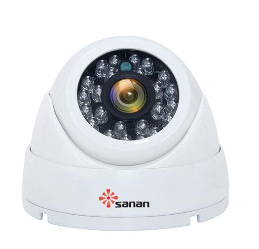
We may have heard of the Dome Fixed Focus Camera/ Fixed Focus Dome Camera/ Fixed Focus Dome cameras/ Fixed Focus Digital Dome Camera/ Fixed Focus Dome Camera Amazon/ Fixed Focus Dome Camera CCTV, so what is the Dome Fixed Focus Camera? We know that the dome refers to the camera shape, so what about the Fixed Focus? Actually, the Fixed Focus refers to a type of the camera lens. Today let me introduce the camera lens for you.
Last time, I have introduced the camera lens and one classification for you. Today please let me introduce the 2nd classification for you---the classification according to the occasions.
In conclusion, we can choose rake the angle of the view into consideration according to our needs, when we choose a Dome Fixed Focus Camera.
Fixed Focus Dome Camera, Fixed Focus Dome cameras, Fixed Focus Digital Dome Camera, Fixed Focus Dome Camera Amazon, Fixed Focus Dome Camera CCTV SHENZHEN SANAN TECHNOLOGY CO.,LTD , https://www.sanan-cctv.com
1. The standard lens
The lens with the angle of view at about 50°(which is also the angle that a person can see without turning his head and eyes), is called the standard lens. The focal length of the 5mm camera's standard lens is mostly 40mm, 50mm or 55mm. The focal length of the 120 camera's standard lens is mostly 80mm or 75mm. The larger the CCD chip is, the longer the focal length of the standard lens will be.
2. The wide-angle lens
The wide-angle lens has the angle of view at above 90 degrees, which is suitable for shooting close and large-scale scenery, and can deliberately exaggerate the foreground to show a sense of perspective. The typical wide-angle lens of the 35mm camera has a focal length at 28mm and an angle of view at 72°. The 50mm, 40mm lens of the 120 camera is equivalent to the 35mm, 28mm lens of the 35mm camera.
3. The long focal length lens
The long focal length lens is suitable for shooting distant subjects. The small depth of field can easily make the background blurred and the subject stand out, but it is bulky and the difficult to focus on the dynamic subjects. The long focal length lenses of the 35mm camera are usually divided into three levels, 135mm or less is called the medium focal length, 135-500mm is called the long focal length, and above 500mm is called the super long focal length. The 150mm lens of the 120 camera is equivalent to the 105mm lens of the 35mm camera. The long focal length lens has the telephoto lens design because it is too bulky, that is, a negative lens is added to the lens behind , and the main plane of the lens is moved forward, then a shorter lens body can be used to obtain the long focal length effect.
4. The reflective telescope lens
The reflective telescope lens is another design of the super telescope lens, which uses a reflecting mirror to form an image. However, due to the design, the aperture can't be installed, and the exposure can be adjusted only through the shutter.
5. The Macro lens
The Macro lens can not only do the close-up macro photography, but also do the telephoto.
