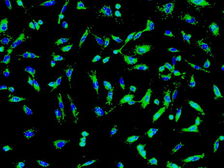
Human umbilical vein endothelial cells (No. #8000 , HUVEC) of Sciencell Company are endothelial cells commonly used in scientific research experiments. When human umbilical vein endothelial cells reach 80-90% fusion, human umbilical vein endothelial cells need to be subcultured. Subculture method: 1. Preparation of the culture flask: The culture flask was coated with fibronectin (Cat . No. #8248 , BPF) at 2 ug/cm2, and the coating time was 37 degrees one hour or 4 degrees overnight. The medium (Cat. #1001 , ECM) , trypsin neutralization solution (Cat . #0113 , TNS) , trypsin/EDTA digest (##103, T/S) and phosphate buffer without calcium and magnesium ions ( Item No. #0303 , DPBS) , the temperature is returned to room temperature, we do not recommend heating the reagent and medium with a 37 ° C water bath. 3. Terminate digestion: Place the culture flask in a 37-degree incubator for 1-2 minutes. At the end of the incubation period, gently pat the flask to remove the umbilical vein endothelial cells from the wall. Morphological changes of umbilical vein endothelial cells were observed under a microscope until 80% of umbilical vein endothelial cells became round. Immediately add 10 ml of trypsin neutralization solution and gently shake the flask. 4. Cell collection: Human umbilical vein endothelial cells were harvested and transferred to a 50 ml centrifuge tube. The culture flask was rinsed by adding 10 ml of medium to ensure that all residual umbilical vein endothelial cells were collected. The number of remaining umbilical vein endothelial cells in the culture flask was observed under a microscope to determine whether most of the umbilical vein endothelial cells were collected, and the number of remaining umbilical vein endothelial cells should be less than 5%. 5. Centrifugal resuspension: The umbilical vein endothelial cells were centrifuged at 1000 rpm (1000 rpm) for 5 minutes, the supernatant was discarded, and the umbilical vein endothelial cells were resuspended in the newly added medium. 6. Plate culture: Count the umbilical vein endothelial cells, and then inoculate them into a new fibronectin-coated culture flask, and culture at 37 degrees. Anesthesia Medical Co., Ltd. , https://www.trustfulmedical.com
2. Trypsinization : Discard the medium in the original culture flask, rinse human umbilical vein endothelial cells with 10 ml of DPBS, and then add 2 ml of trypsin digest . Gently shake the flask until the trypsin digest contains all human umbilical vein endothelial cells. If the pancreatin/EDTA digest of ScienCell Laboratories is used, the damage to umbilical vein endothelial cells will be minimized by trypsinization.