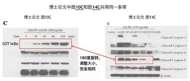
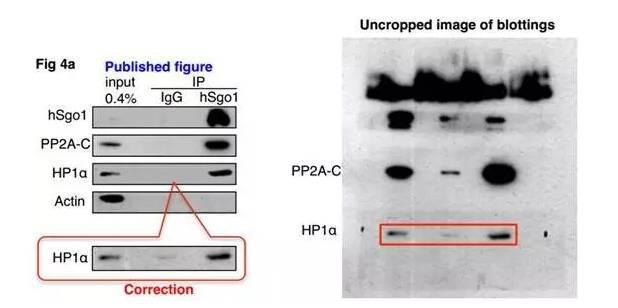
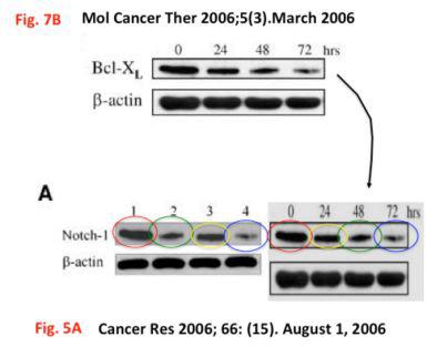
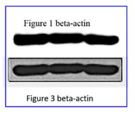
Recent academic news about western blot data fraud has become a hot search term for scientific research. Its data falsification includes the use of a multi-image, using cut, splicing, flipping, rotating, resizing, deliberately smoothing the background and other P-pictures. Techniques, let's take a look at the data of these frauds. One, one picture dual use - use the same strip, P picture represents different experimental results. Two different experiments, the same band represents different proteins (GST-IkBa and Caspase) Effects of zinc finger protein A20 on oxidized low density lipoprotein-mediated proliferation and apoptosis of macrophages and vascular smooth muscle cells and its molecular mechanism, June 2005 Second, smooth the background In order to achieve the desired experimental results, intentionally adjust the contrast and erase the unwanted strips. Kagami, A. et al. Acetylation regulates monopolar attachment at multiple levels during meiosis I in fission yeast. EMBO Rep. 12, 1189–1195 (2011). Third, the puzzle paste ABCD becomes ACBD --Metformin inhibits cell proliferation, migration and invasion by attenuating CSC function mediated by deregulating miRNAs in pancreatic cancer cells. Cancer Prevention Research (2012) Fourth, a group of controls used multiple times Like GAPDH and β-actin, these are basically positive results, so sometimes some small partners can save time and do a GAPH, β-actin WB map, you can use a while, send a few articles, sometimes tune Brightness, sometimes the following contrast is increased, and sometimes the image is reversed. --Increased Ras GTPase activity is regulated by miRNAs that can be attenuated by CDF treatment in pancreatic cancer cells.Cancer Letters (2012) So how do we curb the occurrence of such fraudulent data, the best way is to choose appropriate and reasonable detection methods and provide real picture information. A and z testers should use the multi-fluorescence western blot method with wider dynamic range to perform western blot detection. The multiplex fluorescence detection secondary antibody uses fluorophore labeling, and the target protein concentration on the membrane is linear with the fluorescence signal intensity. To achieve true quantitative requirements, without the need for a glass peeling membrane and secondary incubation, the quality control of the sample, the target protein and the internal reference protein on the same membrane, the above data fraud will not occur. In summary, to choose the appropriate western blot detection method, good imaging detector and a positive academic heart, we can do scientific research without similar events. The Azure C Series multi-function imaging system and the Azure Sapphire dual-mode multispectral laser imaging system escort your experimental data. Medical Caps,Head Cap Medical,Medical Head Cap,Disposable Medical Caps Xinxiang Huaxi Sanitary Materials Co., Ltd. , https://www.huaximedical.com


