High-content technical advantage The high-content cell imaging analysis system consists of three parts: fully automated high-speed microscopy, fully automated image analysis and data management. Fully automated high-speed microscopy produces a large number of images in a short period of time. Fully automated image analysis extracts large amounts of data from these images. Data management software is responsible for document storage, annotation comparison, and retrieval of these images and data. High content means rich information. This information includes: individual cell images and indicators, statistical analysis of cell populations, changes in cell number and morphology, changes in subcellular structure, changes in fluorescence signals over time, changes in the spatial distribution of fluorescent signals, and so on. People often design experiments for specific problems, and while finding answers in the images, other information can lead to unexpected new discoveries. Fluorescence microscopy images of a single sheet in scientific papers usually show the presence or absence of a positive target, strong or weak, and are qualitative analyses. The research object of high-content technology is not one or a few cells, but the simultaneous observation and analysis of a large number of cells, and the quantitative statistical results of cell populations are obtained from a large number of images, and the conclusion is more scientific and persuasive. Because the object of analysis is intact cells, multiple fluorescent probes can be used simultaneously in one experiment, and multi-parameter simultaneous analysis of a large number of cells provides a deeper understanding of complex cytological mechanisms and interactions. High-intensity technology enables high-speed microscopic imaging, which was previously completed in a time-consuming and labor-intensive process, and combined with massive information to make decisions faster. In confirmation of drug targets, pharmacology and toxicity screening compounds HCA obvious advantages for the study of complex cellular mechanisms, the valuable information provided by HCA can help researchers to win in the increasingly fierce competition in scientific research. High-content technology eliminates human bias, people choose better cells for shooting and analysis, and the machine is objective, and the imaging analysis conditions of all samples are identical. High content technology application High-content technology was first applied to drug screening . With the advancement and development of technology, it has been widely used in life science research in recent years, including cell signaling pathways, oncology, neurobiology, immunology, infectious diseases, stem cells. Research and so on. The information provided by HCA cannot be obtained from traditional methods. Many experiments are done with high-content platforms, which are more sensitive than existing methods, with higher throughput, lower cost, and more accurate and reliable results. RNAi technology can specifically eliminate or turn off the expression of specific genes, so this technology has been widely used to explore gene function, into many fields of biological science, and become a major biological research tool. A number of reports have reported the use of high-content platform high-throughput analysis of cell phenotypic changes for RNAi screening to find target genes, and IN Cell users have published nearly 30 such papers. Zebrafish is used as a model organism for genetics and developmental biology research. It is also widely used in drug screening, and has established many zebrafish models of human major diseases such as nervous system diseases, tumor angiogenesis, and congenital heart disease. The zebrafish embryos are transparent and can be viewed under a microscope without fixation. The small volume can be cultured in 96 or 384-well plates for easy application of various conditions for high-throughput screening. When the IN Cell 2000 large-chip CCD camera is combined with a 2× objective lens, it can image the entire hole of the 96-well plate. It is especially suitable for the observation of zebrafish embryos. A large number of live zebrafish embryo images can be obtained in a short time. IN Cell Investigator image analysis The Zebra Fish module of the software automatically analyzes these images. An article by Zhejiang University X.Xu et al. is published in J Neurosci Methods. 2011 Sep 15;200(2):229-36. 96 hole single hole Digital analysis of various organs of zebrafish embryos Gantry Type Video Measuring Instrument
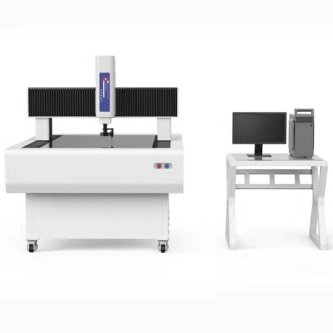
The gantry image measuring instrument can efficiently detect the contours, surface shapes, dimensions, angles, and positions of various workpieces. It is mainly used to measure the size and angle of parts and components that are difficult or impossible to measure with caliper and angle ruler, but play an important role in assembly. It can also be used to take pictures of some parts and components to analyze the causes of defects, especially for PCB boards, films, protective films, optical glass, electronics, large sheet metal parts, connectors, precision mechanical parts, electronic components, semiconductor components and other industries, It can achieve rapid and accurate detection in large quantities.
advantage: Gantry Type Video Measuring Instrument,Large Image Measuring Instrument,Large Video Measuring Instrument,Video Inspection System Zhejiang dexun instrument technology co., ltd , https://www.dexunmeasuring.com

1. Adopting a high-power industrial high-definition color CCD and a continuous automatic zoom depth lens with fixed frame position, the zoom rate does not require calibration, greatly improving work efficiency and measurement accuracy.
2. Images can be output to electronic computer software for surveying and mapping engineering storage. It can be used for photography, copying, and storage.
3. It can inspect the specifications of buried holes, pipe trenches, etc. on the upper surface of the tested object block. The image and color tone on the surface of the product workpiece can be clearly seen.